New ultrathin optical device can precisely capture and stimulate the mammalian brain
Reliably tracking and manipulating the mammalian nervous system in laboratory or clinical settings allows neuroscientists to test their hypotheses, which may in turn lead to new important discoveries. The most well-established and widely used technologies for studying the brain utilize electrodes, devices that can monitor or stimulate electrical activity in their surroundings.
Yet recent studies on mice, non-human primates and other mammals have also highlighted the promise of optical and optogenetic techniques for studying the activity of neurons in the mammalian brain. The advantage of optical techniques is that they can target specific neuron populations with high levels of precision, at greater distances and spanning across larger cortical areas, allowing neuroscientists to meticulously track and modulate neural activity.
Despite their potential, these techniques typically rely on the use of bulky and sophisticated lab instruments, such as tabletop microscopes. Some computer scientists and engineers have tried introducing less bulky and more affordable solutions, such as lensless miniature microscopes that capture and digitally reconstruct images by performing computations. Yet even these solutions have limitations, such as lower resolutions than lens-based optical techniques and greater computational requirements.
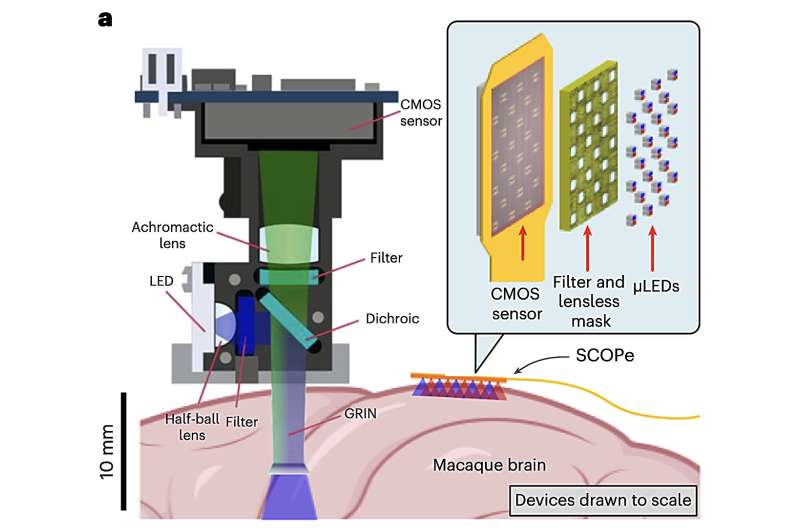
Researchers at Columbia University, New York University and other institutes recently developed a new subdermal optical device that could be used to monitor and stimulate the brain with greater precision. This device, introduced in a paper in Nature Electronics, relies on a complementary metal-oxide semiconductor (CMOS)-based optical probe.
“There has been considerable progress in miniaturizing microscopes for head-mounted configurations, but existing devices are bulky and their application in humans will require a more non-invasive, fully implantable form factor,” wrote Eric H. Pollmann, Heyu Yin and their colleagues in their paper. “We report an ultrathin, miniaturized subdural CMOS optical device for bidirectional optical stimulation and recording.”
The optical probe that the team’s device is based on, called SCOPe, is comprised of a flexible, lens-less and thin miniature microscope, as well as an optical stimulator. Notably, the probe is thin enough to fit in the subdural space of a primate’s brain; a narrow area between two layers of tissue that cover the mammalian brain, known as the dura mater and arachnoid mater.
“We use a custom CMOS application-specific integrated circuit that is capable of both fluorescence imaging and optogenetic stimulation, creating a probe with a total thickness of less than 200 µm, which is thin enough to lie entirely within the subdural space of the primate brain,” wrote Pollmann, Yin and their colleagues. “We show that the device can be used for imaging and optical stimulation in a mouse model and can be used to decode reach movement speed in a non-human primate.”
As part of their study, the researchers tested their device on mice, successfully demonstrating its promise for both imaging and optically stimulating the mouse brain. Subsequently, they also used their device to study the activity of neurons in the motor cortex of non-human primates.
The results gathered in their initial tests were highly promising, as the device allowed them to image the whole brain region of interest, while also allowing them to correlate the animals’ movements with brain activity. In the future, this new promising technology could open interesting possibilities for research, allowing other neuroscientists to precisely manipulate and monitor the activity of specific neurons in a less-invasive way within the brains of animals as they are engaged in specific activities.
More information:
Eric H. Pollmann et al, A subdural CMOS optical device for bidirectional neural interfacing. Nature Electronics(2024). DOI: 10.1038/s41928-024-01209-w
© 2024 Science X Network
Citation:
New ultrathin optical device can precisely capture and stimulate the mammalian brain (2024, October 6)
retrieved 7 October 2024
from https://techxplore.com/news/2024-10-ultrathin-optical-device-precisely-capture.html
This document is subject to copyright. Apart from any fair dealing for the purpose of private study or research, no
part may be reproduced without the written permission. The content is provided for information purposes only.

Comments are closed